I have to create some 3D illustration of bacterias, using a microscope look. See below the samples I have to model from.
TomG created some shaders for ShaderLab that could work for the right and middle picture, but I wonder how to recreate the look of the first picture on the left. A Photoshop filter rather than tS shader perhaps ?
Any idea someone ?
ShaderLab was created in the tS4/5 years ; did anyone tried to achieve a better look with our current tS7.6 modeler or workspace tools ?
RAYMAN
Captain
Posts:2547
Joined: 21 May 2009, 18:56
Re: Scanning Electrons Microscope shader
Postby RAYMAN »
Emmanuel wrote:I have to create some 3D illustration of bacterias, using a microscope look. See below the samples I have to model from.
TomG created some shaders for ShaderLab that could work for the right and middle picture, but I wonder how to recreate the look of the first picture on the left. A Photoshop filter rather than tS shader perhaps ?
Any idea someone ?
ShaderLab was created in the tS4/5 years ; did anyone tried to achieve a better look with our current tS7.6 modeler or workspace tools ?
Illus3D.jpg
The first one is probably best made from a glass shader with a bump and the other two are very good examples of subsurface scattering effects with a dull surface sometimes also called a bloodcell shader...
in a package like Vue infinite you can have the surface of an object have a different sub surf look then the center via the
node based system....
I remember and recall there was the same thing from symbiont- darktree for the Truespace render engine... http://www.darksim.com/html/download_simbiont2.shtml"
Emmanuel
Chief Warrant Officer
Posts:670
Joined: 14 Jun 2009, 06:47
Re: Scanning Electrons Microscope shader
Postby Emmanuel »
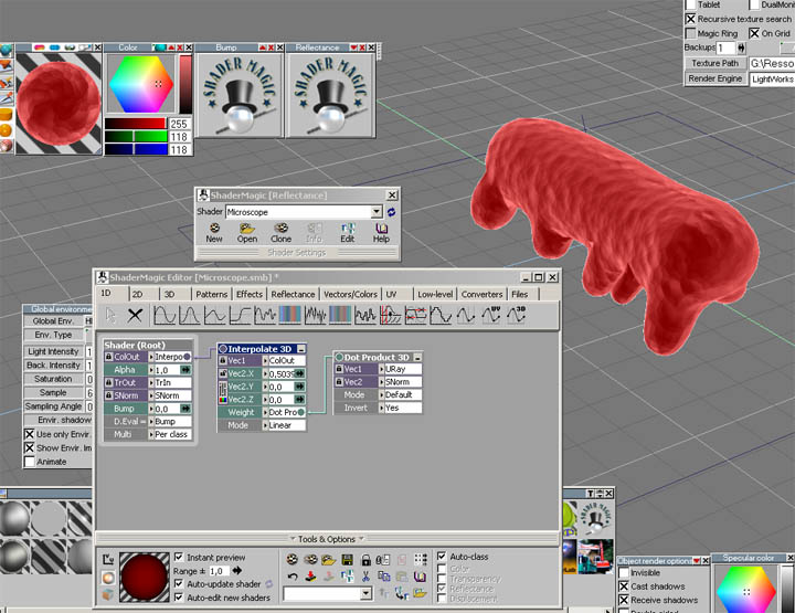
I just found a Microscope shader in ShaderMagic. It seems to do the trick... just have to complete the scene and tweak the shader settings and light setup
Thank you for your input Rayman!
RAYMAN
Captain
Posts:2547
Joined: 21 May 2009, 18:56
Re: Scanning Electrons Microscope shader
Postby RAYMAN »
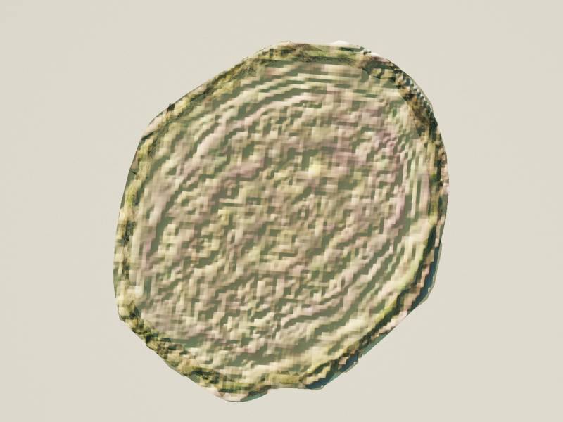
I used your map and a transparany with a bump to get the first image model....
here you go...
Peter
Attachments
Emmanuel
Chief Warrant Officer
Posts:670
Joined: 14 Jun 2009, 06:47
Re: Scanning Electrons Microscope shader
Postby Emmanuel »
Yes! A bump with texture map from the picture, and a glass shader... here it is !
Thanks Rayman
clintonman
Captain
Posts:5659
Joined: 21 May 2009, 21:08
Type the number ten into the box: 0
Location: California
Re: Scanning Electrons Microscope shader
Postby clintonman »
RAYMAN wrote:I used your map and a transparany with a bump to get the first image model....
here you go...
Peter
Kind of looks like a potato chip. Hmm, maybe I should go eat something.
[Disclaimer: the following may sound more natural if your mind's ear "hears" it in the voice of Professor John I.Q. Nerdelbaum Frink from "The Simpsons" and if you preface random sentences with an adenoidal "Nyh'aAAac-tually..."]
In the interest of scientific accuracy, I ought to mention that the second and third sample images do appear to have been made by a scanning electron microscope, then hand-colored (electron microscopes don't "see" in color), the first appears to be from a traditional light-based transmission microscope -- the kind you think of when you read the word "microscope," the kind you can actually buy for a few hundred dollars and use in any home, lab, or school.
SEMs require a solid specimen -- if ordinarily liquid, it must be frozen -- and can view only its surface and immediately below. They do this by scanning a beam of electrons over the surface and picking up what bounces off (there are several possibilities; a given SEM won't detect all of them but it'll be enough), then building an image from the varying signal returned by bounce / x-ray emission / what-have-you.
Any optical characteristics or effects, such as transparency, color, luster; chromatic aberration, depth of field, etc. aren't picked up / don't occur, since it's not light but electrons that are used. Everything is very sharp; no focusing is required, as you aren't dealing with a pencil of wild photons but a controlled beam of quite domesticated electrons.
There are also Transmission Electron Microscopes, which pass an electron beam through the specimen. For electrons to penetrate and come out the other side the specimen must be an incredibly thin slice, a fraction of a nanometer or so. But I don't think any of those pictures come from one.
*removes white lab coat and thick spectacles* *puts down clipboard*